Mrt Cranial
This MRI brain cross sectional anatomy tool is absolutely free to use Use the mouse scroll wheel to move the images up and down alternatively use the tiny arrows (>>) on both side of the image to move the images>>) on both side of the image to move the images.
Mrt cranial. Magnetic resonance angiography – also called a magnetic resonance angiogram or MRA – is a type of MRI that looks specifically at the body’s blood vessels. And 35 MRT cases (19 RTKs, 12 extrarenal MRTs, and 4 cases from unknown tissue types) Nearly all (31/32) ATRTs in this group were classified as ATRTMYC The ‘‘RTKlike’’ Group 3 (n = 59) consisted of 2 ATRT and 57 MRT cases, of which 53 MRT cases were RTKs The ‘‘extrarenal MRTlike’’ Group 4. UPRIGHT MultiPosition MR scanning has uncovered a key set of new observations regarding Multiple Sclerosis (MS), which observations are likely to provide a new understanding of the origin of MS The new findings may also lead to new forms of treatment for MS The UPRIGHT MRI has demonstrated pronou.
Magnetic resonance imaging (MRI) of the head is a painless, noninvasive test that produces detailed images of your brain and brain stem An MRI machine creates the images using a magnetic field and. Full labeled MRI Normal anatomy of the cervical spine (cervical vertebrae) using crosssectional (axial, sagittal and coronal) magnetic resonance where the vertebrae, the nervous system, the intervertebral discs and the zygapophyseal joints and the vascularization can be differentiated This imaging was created from sagittal T1weighted sequences and T2 reconstructions. Discussion IIH, also known as pseudotumor cerebri and benign intracranial hypertension, is a syndrome characterized by increased CSF pressure and papilledema in patients without focal neurologic findings, except for occasional CN VI palsy It is a diagnosis of exclusion, and radiologic examinations are traditionally performed to help exclude lesions that produce intracranial hypertension.
The MRT's capabilities are proving especially valuable for procedures in the head and neck, said neuroradiologist Dr Alex Norbash, an assistant professor of radiology such as cranial nerves. Magnetic Resonance Imaging (MRI) is a commonly accepted and widely used diagnostic medical procedure It is often safe to perform MRI on an individual that has an orthopaedic implant device. The conclusion of a recent large cohort study from Ontario, Canada (Ray JG et al JAMA 16;316(9)) states, "Exposure to MRI during the first trimester of pregnancy compared with nonexposure was not associated with increased risk of harm to the fetus or in early childhood Gadolinium MRI at any time during pregnancy was associated with an increased risk of a broad set of.
There is mounting evidence that cranial nerve involvement in COVID19 represents autoimmunity, as in GBS cases 2 First, GBS is the prototypical postviral induced neuropathy seen in 70% of cases with known triggers, including influenza, enteroviruses, H1N1, West Nile virus, Zika, MERSCoV, and SARSCoV 7 Second, in 7 of 11 tested patients with. This MRI brain cross sectional anatomy tool is absolutely free to use Use the mouse scroll wheel to move the images up and down alternatively use the tiny arrows (>>) on both side of the image to move the images>>) on both side of the image to move the images. Brain imaging, magnetic resonance imaging of the head or skull, cranial magnetic resonance tomography (MRT), neurological MRI they describe all the same radiological imaging technique for.
Cranial ultrasound (CUS) is a reputable tool for brain imaging in critically ill neonates Methods MRT/CT images from 117 children with hydrocephalus were evaluated at time of diagnosis. Region specific anatomic shape for efficient and aesthetic placement. Hardbound MRI Textbook MRI BIOEFFECTS, SAFETY, AND PATIENT MANAGEMENT is a comprehensive, authoritative textbook on the health and safety concerns of MRI technology that contains contributions from more than forty internationally respected experts in the field It serves as the definitive resource for radiologists and other physicians, MRI technologists, physicists, scientists, MRI facility.
Overview Whiplash is a neck injury due to forceful, rapid backandforth movement of the neck, like the cracking of a whip Whiplash is commonly caused by rearend car accidents. (c) "foreends", for the purposes of subheadings 03 19 11, 03 29 11, 0210 19 30 and 0210 19 60 the anterior (cranial) part of the halfcarcase without the head, with or without the chaps, including bones, with or without foot, shank, rind or subcutaneous fat. Localized cranial arterial involvement could be detected due to inflammatory signal enhancement in the superficial left occipital artery (Fig 3, postcontrast) The T 1 contrast images clearly reveal signal enhancement due to the accumulation of contrast agent and circumferential luminal thickening as visible for the magnified right (bottom.
This section of the website will explain large and minute details of venous anatomy of brain. Hier erklären Ärzte leicht verständlich Begriffe aus medizinischen Befunden. Twentytwo magnetic resonance imaging (MRI) brain studies of different breeds of dogs were reviewed to assess the anatomy of cranial nerve (CN) origins and associated skull foramina These included five anatomic studies of normal brains using 2mmthick slices and 17 studies using conventional clini.
Cranial ultrasound (CUS) is a reputable tool for brain imaging in critically ill neonates Methods MRT/CT images from 117 children with hydrocephalus were evaluated at time of diagnosis. And release of the retrohyoid fascia More information about the clinical protocol is given by RodríguezFuentes et al 17. For these reasons, CISS sequence is very useful for evaluating structures surrounded by CSF (eg cranial nerves) MRI image appearance The easiest way to identify CISS images is to look for fat and fluid filled space in the body (eg cerebrospinal fluid in the brain ventricles and spinal canal) Fluids normally appear as very bright and fat.
An atypical teratoid rhabdoid tumor (AT/RT) is a rare tumor usually diagnosed in childhood Although usually a brain tumor, AT/RT can occur anywhere in the central nervous system (CNS), including the spinal cordAbout 60% will be in the posterior cranial fossa (particularly the cerebellum)One review estimated 52% in the posterior fossa, 39% are supratentorial primitive neuroectodermal tumors. CNS = brain, brain meninges, cranial nerves & spinal cord PNS = nerves and ganglia Has both sensory and motor function Sensory send impulses to brain and spinal cord to skeletal muscle Autonomic conducts impulses from brain and spinal cord to smooth muscle tissue Ex Digestive and cardiopulmonary systems. Localized cranial arterial involvement could be detected due to inflammatory signal enhancement in the superficial left occipital artery (Fig 3, postcontrast) The T 1 contrast images clearly reveal signal enhancement due to the accumulation of contrast agent and circumferential luminal thickening as visible for the magnified right (bottom.
This MRI cranial nerves axial cross sectional anatomy tool is absolutely free to use Use the mouse scroll wheel to move the images up and down alternatively use the tiny arrows (>>) on both side of the image to move the images>>) on both side of the image to move the images. Malignant rhabdoid tumors (MRTs) are rare lethal tumors of childhood that most commonly occur in the kidney and brain MRTs are driven by SMARCB1 loss, but the molecular consequences of SMARCB1 loss in extracranial tumors have not been comprehensively described and genomic resources for analyses of extracranial MRT are limited. This cover page design template is complete compatible with Google Docs Just download DOCX format and open the theme in Google Docs Unfortunately, the item MRT Of Cranial Cavity Word Template id which price is Free has no available description, yet.
Intraparenchymal cerebral cavernous malformation is difficult to localize intraoperatively with conventional frameless navigation due to the “brain shift” effect We conducted this study to evaluate the efficacy and safety of intraoperative magnetic resonance image (iMRI)assisted neuroport surgery for the resection of cerebral intraparenchymal cavernous malformation. CranioCurve Preformed Mesh is a titanium cranioplasty solution and is part of our comprehensive cranioplasty portfolio, for efficient coverage of cranial defects in multiple anatomic regions Features 06mm grade II titanium for a balance of strength and flexibility;. Introduction MRI is the most sensitive imaging method when it comes to examining the structure of the brain and spinal cord It works by exciting the tissue hydrogen protons, which in turn emit electromagnetic signals back to the MRI machine The MRI machine detects their intensity and translates it into a grayscale MRI image Thus, for describing the MRI appearance of the parts of the.
This section of the website will explain large and minute details of arterial anatomy of brain. Discussion IIH, also known as pseudotumor cerebri and benign intracranial hypertension, is a syndrome characterized by increased CSF pressure and papilledema in patients without focal neurologic findings, except for occasional CN VI palsy It is a diagnosis of exclusion, and radiologic examinations are traditionally performed to help exclude lesions that produce intracranial hypertension. MRT is very rare.
Rhaboid tumors that grow outside of the brain are called extracranial malignant rhabdoid tumor, malignant rhabdoid tumor, or MRT MRTs grow and spread to other parts of the body quickly How common are extracranial malignant rhabdoid tumors?. Was bedeutet cranial in einem MRTBefund vom Kopf?. Which settle in the mesenteric veins (5) The adultwormlayeggsthatareexcretedwithstool or urine Different mechanisms of invasion of thebrainhavebeendiscussedtheeggsmay.
The latest breaking news and information for Midland Texas and Permian Basin from the Midland ReporterTelegram. The intention of this article is to alert readers to common problems and pit falls concerning magnetic resonance imaging (MRI) for pathologies of cranial nerves It also provides an introduction to helpful sequences and post process ing techniques. CNS = brain, brain meninges, cranial nerves & spinal cord PNS = nerves and ganglia Has both sensory and motor function Sensory send impulses to brain and spinal cord to skeletal muscle Autonomic conducts impulses from brain and spinal cord to smooth muscle tissue Ex Digestive and cardiopulmonary systems.
Hier erklären Ärzte leicht verständlich Begriffe aus medizinischen Befunden. This section of the website will explain large and minute details of venous anatomy of brain. Release of the suprahyoid and infrahyoid muscles;.
The craniocervical junction (CCJ) is comprised of the inferior surface of the skull, the atlas and axis, as well as muscles and connective tissues that attach the skull to the cervical spine The CCJ encloses the central nervous system (CNS), encephalic vasculature and the cerebrospinal fluid (CSF) system The CCJ spans the brainstem to the spinal cord, including the vascular system as well as. Results Based on our experience with the firstinhuman, neuromodulationencapsulated, cranial implant case (1), our team was again successful in creating the world’s first functional prototype named the LID MRT Upon completion, it required the size dimensions most consistent with a standard hemicraniectomy and fully functions with respect. Twentytwo magnetic resonance imaging (MRI) brain studies of different breeds of dogs were reviewed to assess the anatomy of cranial nerve (CN) origins and associated skull foramina These included five anatomic studies of normal brains using 2mmthick slices and 17 studies using conventional clini.
Was bedeutet cranial in einem MRTBefund vom Kopf?. This cover page design template is complete compatible with Google Docs Just download DOCX format and open the theme in Google Docs Unfortunately, the item MRT Of Cranial Cavity Word Template id which price is Free has no available description, yet The item rating has 46 star(s) with 23 votes. To facilitate comparisons between cranial and extracranial MRTs and to study the genomewide consequences of SMARCB1 loss, we performed comprehensive genomic analyses of pediatric extracranial MRT cases Despite being almost uniformly driven by SMARCB1 loss, we observed molecular heterogeneity within extracranial MRTs.
Region specific anatomic shape for efficient and aesthetic placement. And release of the retrohyoid fascia More information about the clinical protocol is given by RodríguezFuentes et al 17. MRI with contrast enhancement is a valuable tool for detecting and characterizing disease of the cranial nerves Abnormal cranial nerve enhancement on MRI may sometimes be the first or only indication of an underlying disease process.
A PET scan is an imaging exam that’s used to diagnose diseases or issues by looking at how the body is functioning It uses a special dye with radioactive tracers to help the machine capture. Introduction MRI is the most sensitive imaging method when it comes to examining the structure of the brain and spinal cord It works by exciting the tissue hydrogen protons, which in turn emit electromagnetic signals back to the MRI machine The MRI machine detects their intensity and translates it into a grayscale MRI image Thus, for describing the MRI appearance of the parts of the. Cranial CT Scan Medically reviewed by Deborah Weatherspoon, PhD, RN, CRNA A cranial CT scan of the head is a diagnostic tool used to create detailed pictures of the skull, brain, paranasal.
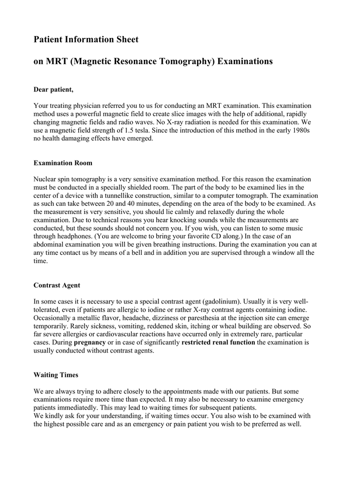
ADVERTISEMENT Radiopaedia is free thanks to our supporters and advertisers Become a Gold Supporter and see no ads. CranioCurve Preformed Mesh is a titanium cranioplasty solution and is part of our comprehensive cranioplasty portfolio, for efficient coverage of cranial defects in multiple anatomic regions Features 06mm grade II titanium for a balance of strength and flexibility;. An MRI scan uses a powerful magnetic field and radio waves to take detailed, 3D pictures of your body The human body is 80 percent water, so it contains millions of hydrogen atoms.
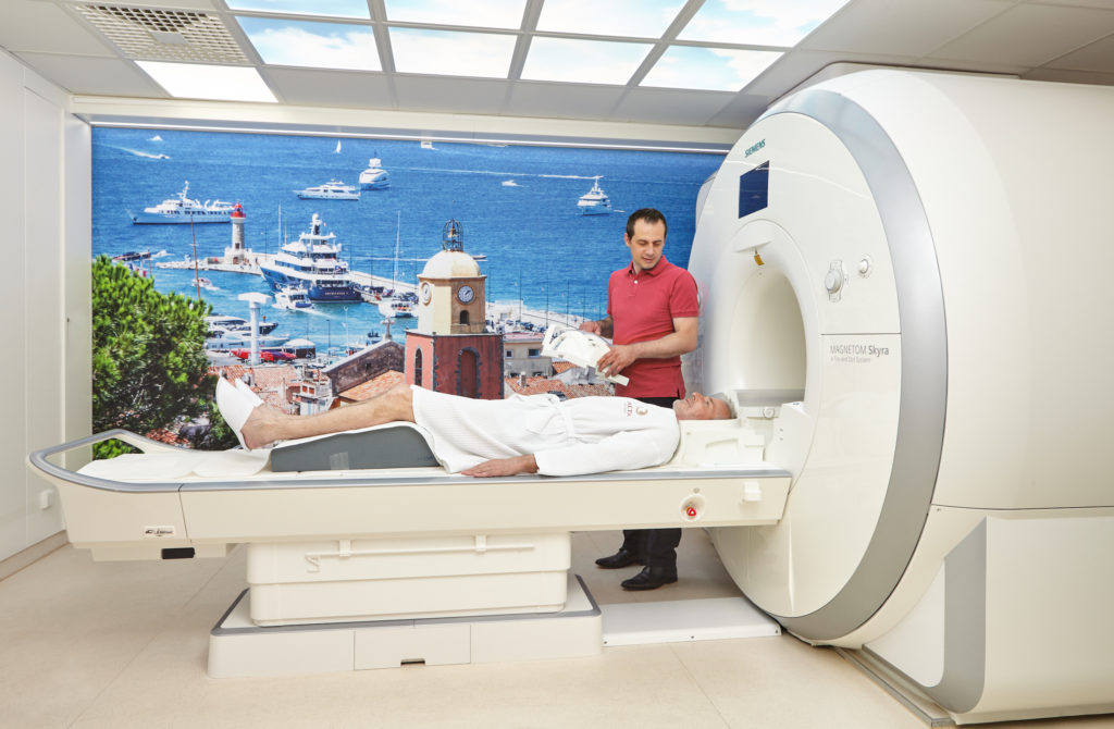
Magnetic resonance imaging (MRI) is a medical imaging technique used in radiology to form pictures of the anatomy and the physiological processes of the body MRI scanners use strong magnetic fields, magnetic field gradients, and radio waves to generate images of the organs in the body MRI does not involve Xrays or the use of ionizing radiation, which distinguishes it from CT and PET scans. A PET scan is an imaging exam that’s used to diagnose diseases or issues by looking at how the body is functioning It uses a special dye with radioactive tracers to help the machine capture. Malignant rhabdoid tumors (MRTs) are rare lethal tumors of childhood that most commonly occur in the kidney and brain MRTs are driven by SMARCB1 loss, but the molecular consequences of SMARCB1 loss in extracranial tumors have not been comprehensively described and genomic resources for analyses of extracranial MRT are limited.
To facilitate comparisons between cranial and extracranial MRTs and to study the genomewide consequences of SMARCB1 loss, we performed comprehensive genomic analyses of pediatric extracranial MRT cases Despite being almost uniformly driven by SMARCB1 loss, we observed molecular heterogeneity within extracranial MRTs Methylation. Brain imaging, magnetic resonance imaging of the head or skull, cranial magnetic resonance tomography (MRT), neurological MRI they describe all the same radiological imaging technique for. The MRT's capabilities are proving especially valuable for procedures in the head and neck, said neuroradiologist Dr Alex Norbash, an assistant professor of radiology such as cranial nerves.
New MRT Imaging Biomarkers and Treatment With Kinetic Oscillatory Stimulation (KOS) in Nasal Cavity for Myalgic Encephalomyelitis/Chronic Fatigue Syndrome (ME/CFS) The safety and scientific validity of this study is the responsibility of the study sponsor and investigators. PURPOSE Malignant rhabdoid tumor (MRT) is a rare and highly aggressive tumor that affects young children Due to its extreme rarity, most of the available data are based on retrospective case series To add to the current knowledge of this disease, we reviewed the patients treated for extracranial MRT in our institute. The MRT involved cranial base release, adjusting the relation of the rectus capitis posterior muscles to the dura mater gross release of the sternocleidomastoid muscle;.
The MRT involved cranial base release, adjusting the relation of the rectus capitis posterior muscles to the dura mater gross release of the sternocleidomastoid muscle;. Release of the suprahyoid and infrahyoid muscles;.

Identification And Analyses Of Extra Cranial And Cranial Rhabdoid Tumor Molecular Subgroups Reveal Tumors With Cytotoxic T Cell Infiltration Sciencedirect

21 Questions With Answers In Mrt Scientific Method
Mrt Scan Of The Cranium Arrow Tumorous Mass In The Left Cellulae Download Scientific Diagram
Mrt Cranial のギャラリー

Magnetic Resonance Imaging Wikipedia

Clinical Cases Fonar Upright Mri

A Global Compound Selectivity Analysis Of Mrt Lines Comparison Of Download Scientific Diagram

Neurovascular Compression At The Ventrolateral Medulla In Autosomal Dominant Hypertension And Brachydactyly Stroke

Virtual Screening Based Discovery And Mechanistic Characterization Of The Acylthiourea Mrt 10 Family As Smoothened Antagonists Molecular Pharmacology
Http Ar Iiarjournals Org Content 40 11 6159 Full Pdf

Usa1 Device For Protecting The Hearing From Loud Mrt Sounds Google Patents

Genome Wide Profiles Of Extra Cranial Malignant Rhabdoid Tumors Reveal Heterogeneity And Dysregulated Developmental Pathways Sciencedirect

A Dynamic Mutational Landscape Associated With An Inter Regionally Diverse Immune Response In Malignant Rhabdoid Tumour Yasui The Journal Of Pathology Wiley Online Library

Genome Wide Profiles Of Extra Cranial Malignant Rhabdoid Tumors Reveal Heterogeneity And Dysregulated Developmental Pathways Abstract Europe Pmc
Grundlagen Der Mrt Und Mrs Springerlink
Plos One Idiopathic Orbital Inflammation Syndrome With Retro Orbital Involvement A Retrospective Study Of Eight Patients

Spinal Mrt A And B Mrt Images Showing The Early Necrosis Of The Download Scientific Diagram

Pdf Mri Based Reevaluation Of Patients With Disc Displacement Without Reduction Mrt Gestutzte Nachuntersuchung Bei Diskusverlagerung Ohne Reposition Semantic Scholar
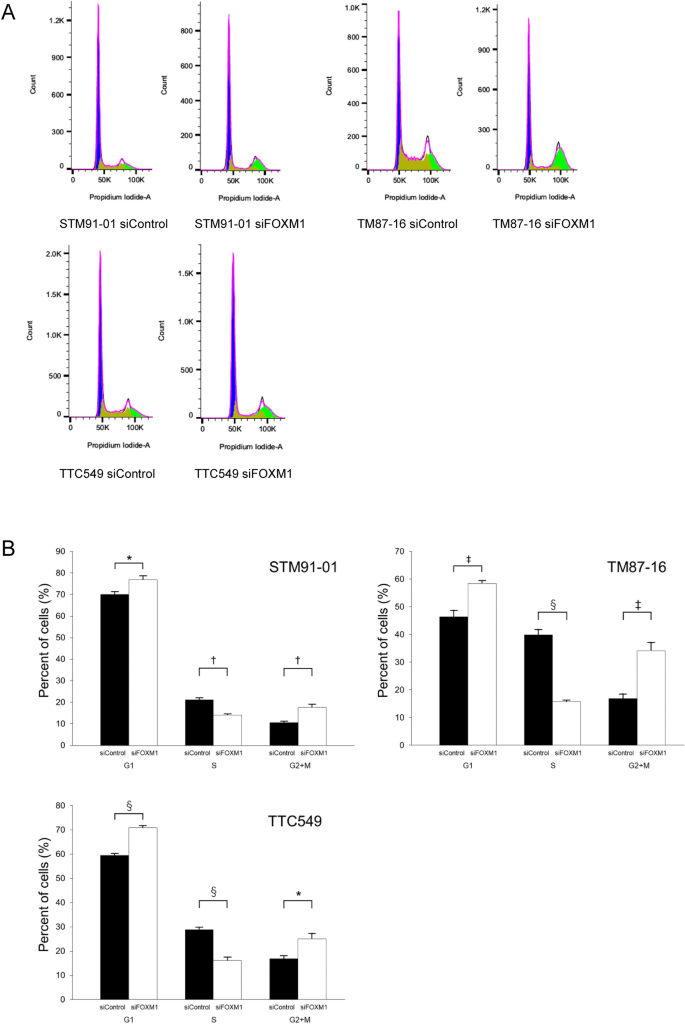
The Forkhead Box M1 Foxm1 Expression And Antitumor Effect Of Foxm1 Inhibition In Malignant Rhabdoid Tumor Springerlink
Mrt Of Cranial Cavity Powerpoint Template Backgrounds 092 Poweredtemplate Com

Identification And Analyses Of Extra Cranial And Cranial Rhabdoid Tumor Molecular Subgroups Reveal Tumors With Cytotoxic T Cell Infiltration Sciencedirect

Identification And Analyses Of Extra Cranial And Cranial Rhabdoid Tumor Molecular Subgroups Reveal Tumors With Cytotoxic T Cell Infiltration Sciencedirect
Www Snec Com Sg Patient Care Conditionstreatments Eye Conditions Brochures Documents En Upper eyelid drooping Pdf

Virtual Screening Based Discovery And Mechanistic Characterization Of The Acylthiourea Mrt 10 Family As Smoothened Antagonists Molecular Pharmacology
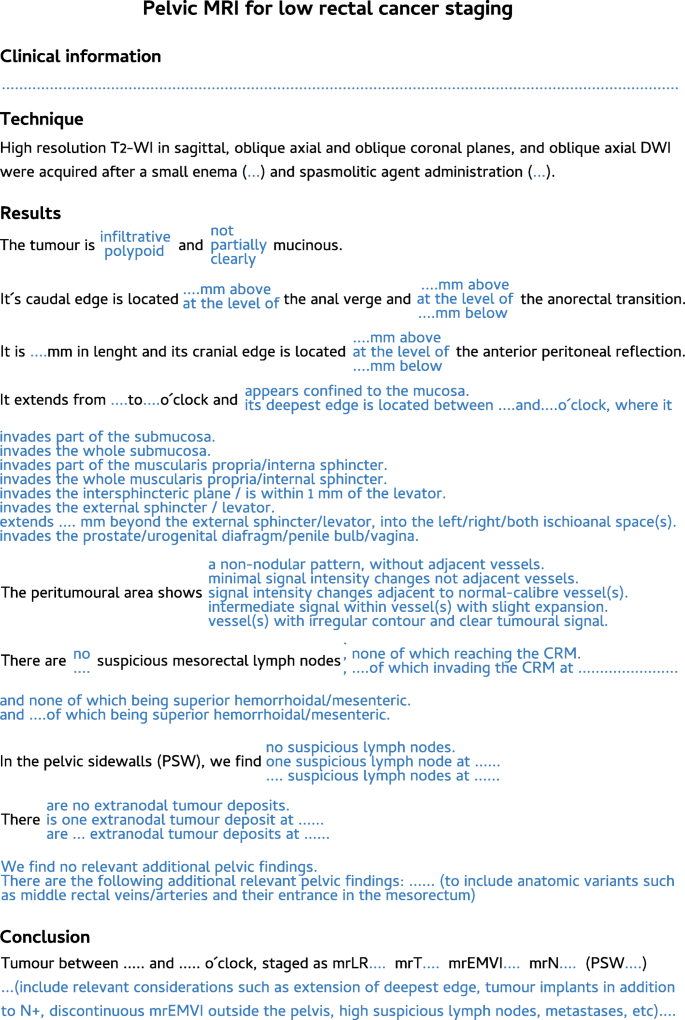
Mri Of Rectal Cancer Relevant Anatomy And Staging Key Points Insights Into Imaging Full Text
Kopf Mrt Ursachen Fur Kopfschmerzen Schwindel Und Druckgefuhl Finden

Novel Two Mrt Cell Lines Established From Multiple Sites Of A Synchronous Mrt Patient Anticancer Research
The Forkhead Box M1 Foxm1 Expression And Antitumor Effect Of Foxm1 Inhibition In Malignant Rhabdoid Tumor Springerlink
Patient Information Sheet On Mrt Magnetic Resonance

Figure 6 From Investigations For Neonatal Seizures Semantic Scholar

File Akustikus Schwannon Rechts Mrt T1km Coronar 001 Jpg Wikimedia Commons

Fmy3xk9ck8aunm

Genome Wide Profiles Of Extra Cranial Malignant Rhabdoid Tumors Reveal Heterogeneity And Dysregulated Developmental Pathways Sciencedirect

Dental Precision Attachments Mrt
2

Improved Outcome Of 131i Mibg Treatment Through Combination With External Beam Radiotherapy In The Sk N Sh Mouse Model Of Neuroblastoma Sciencedirect
Q Tbn And9gctog794rzftwzvgcjzd0fdawd 2fygzisgpiou A6pklkuaqyb5 Usqp Cau

The Measurement Results Of Medial Root Thickness Mm Mrt Lateral Download Scientific Diagram

Clinical Cases Fonar Upright Mri
Mrt Of Cranial Cavity Powerpoint Template Backgrounds 092 Poweredtemplate Com
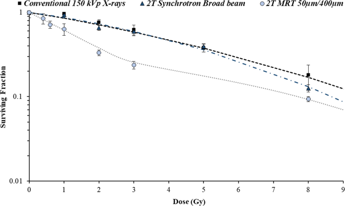
Toward Personalized Synchrotron Microbeam Radiation Therapy Scientific Reports

Identification And Analyses Of Extra Cranial And Cranial Rhabdoid Tumor Molecular Subgroups Reveal Tumors With Cytotoxic T Cell Infiltration Abstract Europe Pmc
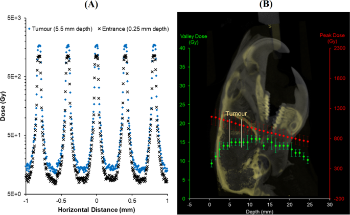
Toward Personalized Synchrotron Microbeam Radiation Therapy Scientific Reports
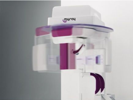
Health Management And Leadership Portal Panoramic X Ray System Dental Radiology Digital Hyperion Mrt Myray Healthmanagement Org

Animal Mrt Cruciate Ligament Tear In Dogs Cruciate Ligament In Dogs Cruciate Ligament Cruciate Ligament Tear
Intracranial Stereotactic Radiosurgery

Identification And Analyses Of Extra Cranial And Cranial Rhabdoid Tumor Molecular Subgroups Reveal Tumors With Cytotoxic T Cell Infiltration Abstract Europe Pmc

Acoustic Neuroma Acoustic Neuroma Operation

Mdm2 And Mdm4 Are Therapeutic Vulnerabilities In Malignant Rhabdoid Tumors Cancer Research
Www Tandfonline Com Doi Pdf 10 1080

Dynamics Of Middle Cerebral Artery Blood Flow Velocity During Moderate Intensity Exercise Abstract Europe Pmc

File Mr Cranial Sinusvene Jpg Wikimedia Commons
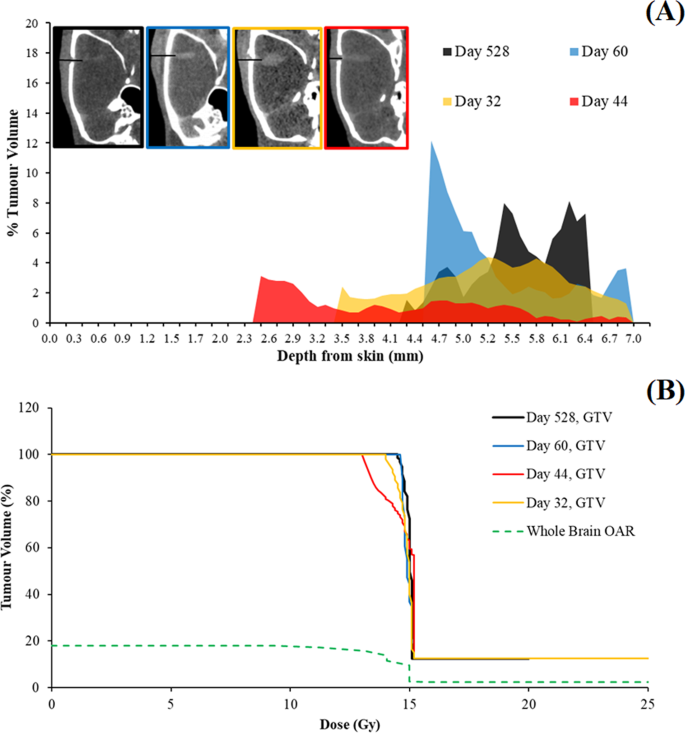
Toward Personalized Synchrotron Microbeam Radiation Therapy Scientific Reports
2

Mdm2 And Mdm4 Are Therapeutic Vulnerabilities In Malignant Rhabdoid Tumors Cancer Research
Q Tbn And9gcs96cjm0kpyqndwbmbfizzqwbk9yzcljggvp8cxihtzd4odv3kx Usqp Cau
Ro Journal Biomedcentral Com Track Pdf 10 1186 S 017 0864 2 Pdf

Figure 6 From Intracranial Hematoma Detection By Near Infrared Spectroscopy In A Helicopter Emergency Medical Service Practical Experience Semantic Scholar
Www Researchsquare Com Article Rs V1 Pdf
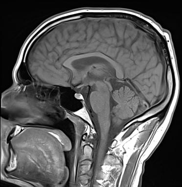
Kopf Mrt Ursachen Fur Kopfschmerzen Schwindel Und Druckgefuhl Finden
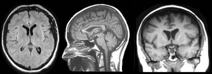
Mri Basics
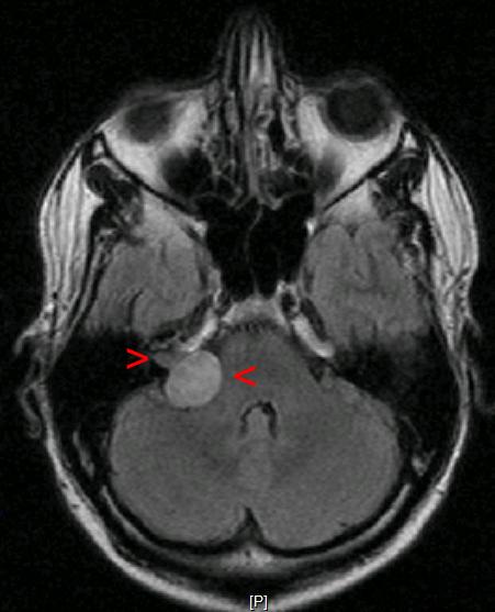
File Akustikusneurinom Mrt Jpg Wikipedia
2
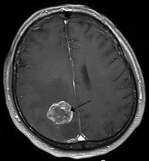
Brain Tumor Wikipedia
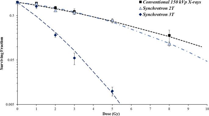
Toward Personalized Synchrotron Microbeam Radiation Therapy Scientific Reports
Http Ar Iiarjournals Org Content 40 11 6159 Full Pdf
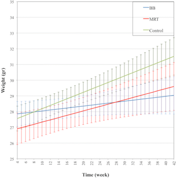
Neurocognitive Sparing Of Desktop Microbeam Irradiation Springerlink

Improved Outcome Of 131i Mibg Treatment Through Combination With External Beam Radiotherapy In The Sk N Sh Mouse Model Of Neuroblastoma Sciencedirect
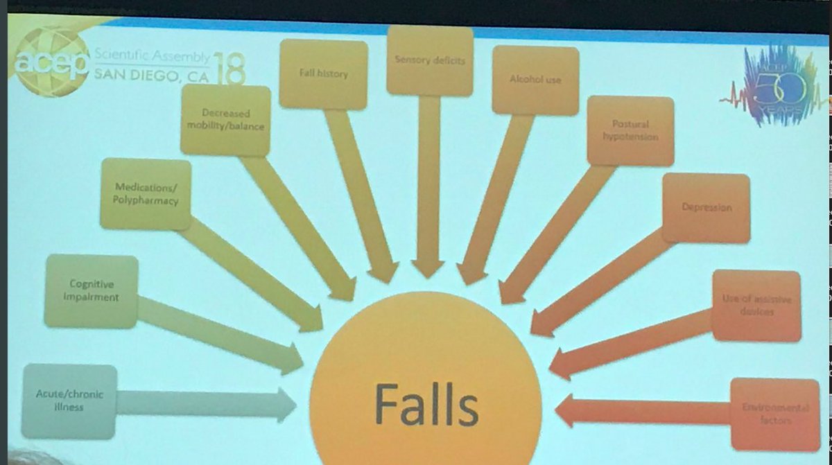
Bernadette Keefe Md Acep18 Via Acep Geried Geriem Geriatricer Mrt Geriatricednews Marcus Escopedo Johnahartford How To Overcome Institutional Inertia Re Geried Adaptation Pending Proof Of Ro Response Begin With

Magnetresonanztomographie Mrt Des Kopfes Ct Mrt Institut Berlin
T2 Weighted Mrt In Coronar Projection Of A Woman With Status After Download Scientific Diagram
Http Ar Iiarjournals Org Content 40 11 6159 Full Pdf

21 Questions With Answers In Mrt Scientific Method
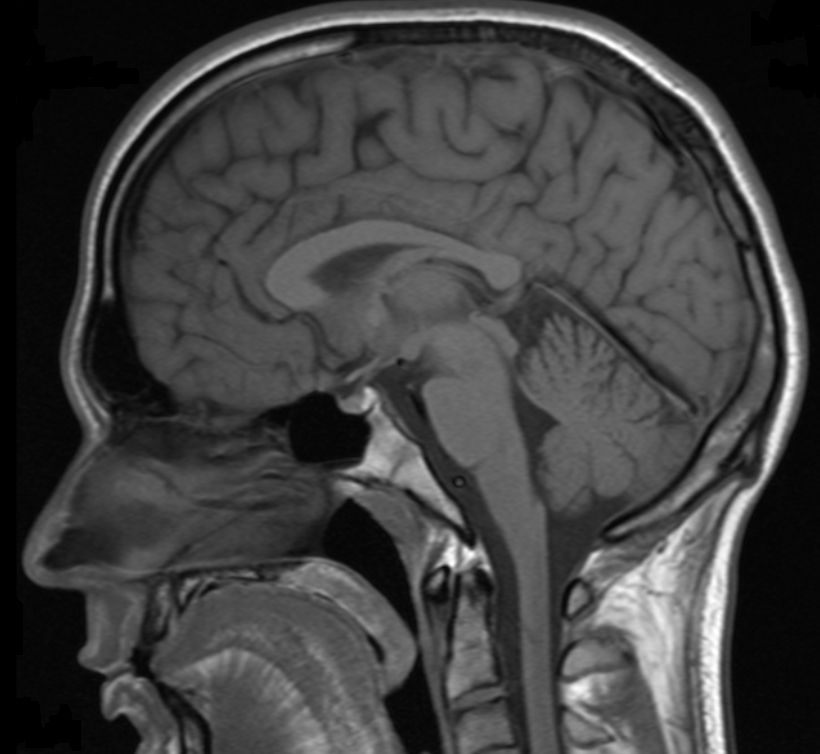
Modelling The Dynamics Of The Brain Medicine Health Information Science Medicine Health Medical Biotechnology Healthcare Delivery V En Science Studentnews Eu

Identification And Analyses Of Extra Cranial And Cranial Rhabdoid Tumor Molecular Subgroups Reveal Tumors With Cytotoxic T Cell Infiltration Sciencedirect
Mrt Of Cranial Cavity Word Template 092 Poweredtemplate Com

Genome Wide Profiles Of Extra Cranial Malignant Rhabdoid Tumors Reveal Heterogeneity And Dysregulated Developmental Pathways Sciencedirect

Clinical Cases Fonar Upright Mri
Www Physicamedica Com Article S11 1797 15 8 Pdf
Osteopathy Podiatry Com Site Wp Content Uploads 18 01 Clinic Brochure Pdf

Cranial Magnetic Resonance Tomography Mrt From Present Patient A T2 Download Scientific Diagram
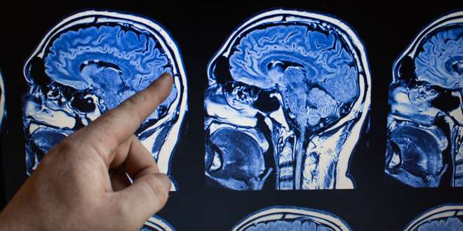
Mrt Des Kopfes

Dual Targeting Of Pdgfra And Fgfr1 Displays Synergistic Efficacy In Malignant Rhabdoid Tumors Sciencedirect

Identification And Analyses Of Extra Cranial And Cranial Rhabdoid Tumor Molecular Subgroups Reveal Tumors With Cytotoxic T Cell Infiltration Sciencedirect

Magnetresonanztomographie Mrt Des Kopfes Ct Mrt Institut Berlin
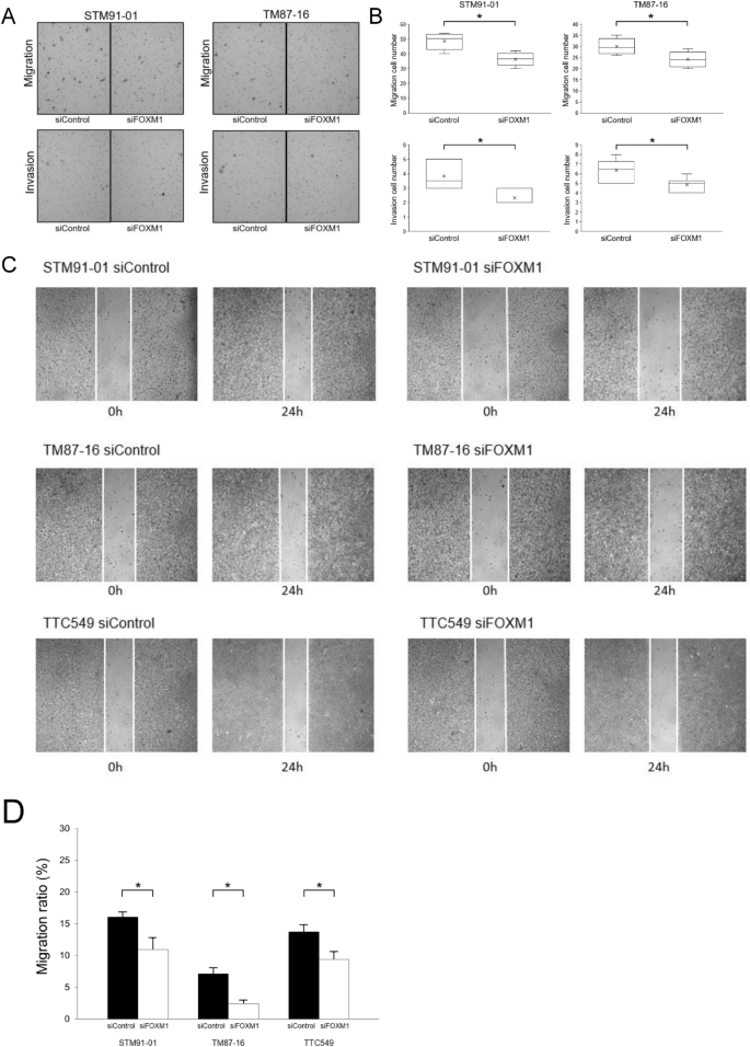
The Forkhead Box M1 Foxm1 Expression And Antitumor Effect Of Foxm1 Inhibition In Malignant Rhabdoid Tumor Springerlink

Brain Mrt Scan Display High Res Stock Video Footage Getty Images

Magnetic Resonance Imaging Wikipedia

Nuklearmedizin Braunschweig Celler Strasse 30 Braunschweig Mrt Des Kopfes

Mrt Of The Brain M H At 7 Years Of Age 11 Depression Of The Download Scientific Diagram
Improved Accuracy Of Breast Volume Calculation From 3d Surface Imaging Data Using Statistical Shape Models
1

Acoustic Neuroma Acoustic Neuroma Operation

Brain Mrt Scan Display High Res Stock Video Footage Getty Images

L M Smyth P A Rogers J C Crosbie J F Donoghue Ppt Download

Acoustic Neuroma Acoustic Neuroma Operation
Www Cell Com Cancer Cell Pdfextended S1535 6108 16 5
Q Tbn And9gcstdwjkagdkwkudbw94 Ou7ssixovcloyittmdcgrhzftv8qbg8 Usqp Cau

Virtual Screening Based Discovery And Mechanistic Characterization Of The Acylthiourea Mrt 10 Family As Smoothened Antagonists Molecular Pharmacology

Genome Wide Profiles Of Extra Cranial Malignant Rhabdoid Tumors Reveal Heterogeneity And Dysregulated Developmental Pathways Sciencedirect
Ro Journal Biomedcentral Com Track Pdf 10 1186 S 017 0864 2 Pdf
Www Cell Com Cancer Cell Pdfextended S1535 6108 16 5

Identification And Analyses Of Extra Cranial And Cranial Rhabdoid Tumor Molecular Subgroups Reveal Tumors With Cytotoxic T Cell Infiltration Sciencedirect

Mrt Kopf Grunde Ablauf Dauer Kosten Praktischarzt

Cranial Magnetic Resonance Tomography Mrt From Present Patient A T2 Download Scientific Diagram


