Major Cardiac Veins
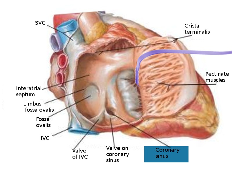
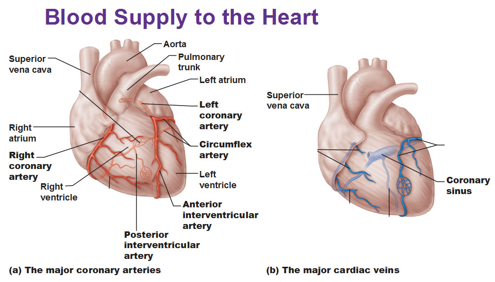
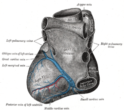
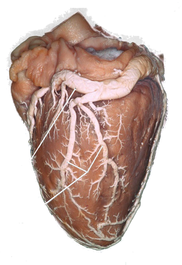
Cardiac Veins Blood travels from the subendocardium into the thebesian veins, which are small tributaries running throughout the myocardiumThese in turn drain into larger veins that empty into the coronary sinus The coronary sinus is the main vein of the heart, located on the posterior surface in the coronary sulcus, which runs between the left atrium and left ventricle.
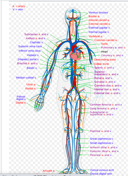
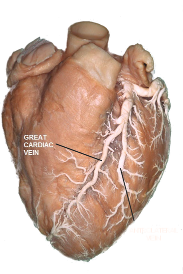
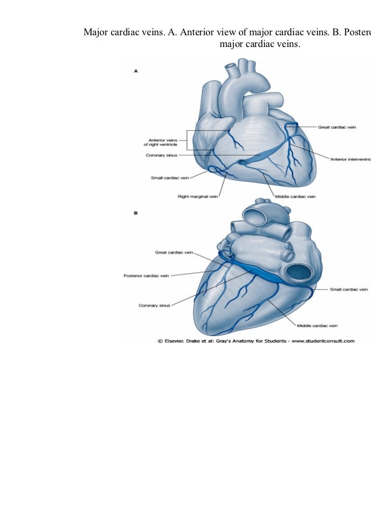
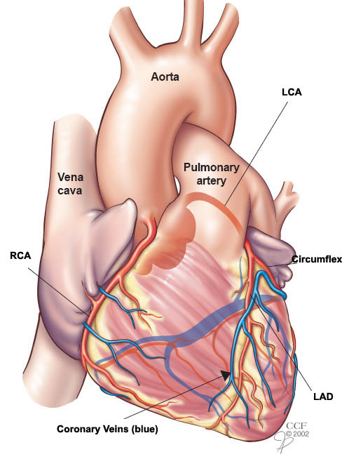
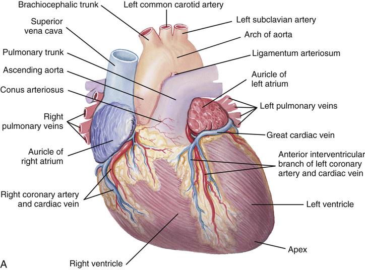
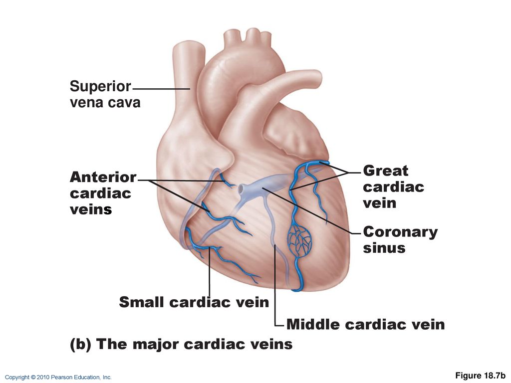
Major cardiac veins. Coronary Veins Coronary veins drain the heart and generally parallel the large surface arteries (see Figure \(\PageIndex{1}\)) The great cardiac vein can be seen initially on the surface of the heart following the interventricular sulcus, but it eventually flows along the coronary sulcus into the coronary sinus on the posterior surface The great cardiac vein initially parallels the anterior. CT cardiac exams provide critical information to practitioners of cardiac therapy Precise information pertaining to the left atrium's complex anatomy, the pulmonary veins, the coronary sinus, or the cardiac veins has a major impact on the efficacy of subsequent cardiac therapy It can speed procedures and facilitate treatment. Passing anterior to the right coronary artery (RCA), the anterior cardiac veins (ACV) commonly enter into the RA as separate vessels or in some cases (5%–27%) form a common trunk The right marginal vein (RMV) is seen in 80% of cases This vein drains into the small cardiac vein (SCV) in 30% of cases and directly into the RA in 70% of cases.
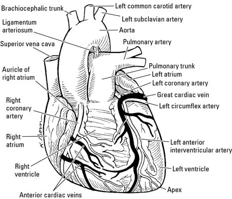
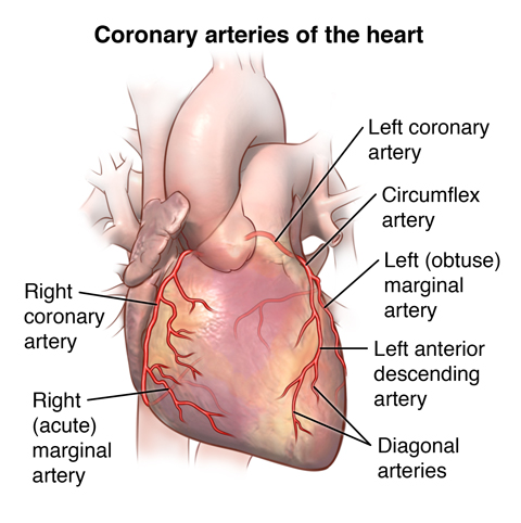
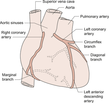
Posterior vein of the left ventricle Anterior cardiac veins Thebesian veins Coronary sinus Great cardiac vein Middle cardiac vein Small cardiac vein Oblique vein of the left atrium Posterior vein of the left ventricle. Cardiac output The volume discharged from the ventricle per minute, calculated by multiplying stroke volume by heart rate, in beats per minute Cardiac veins Those veins that branch out and drain blood from the myocardial capillaries to join the coronary sinus Carotid sinuses Enlargements near the base of the carotid. Here are three branches Anterior interventricular artery Runs along the anterior interventricular groove to the apex;.
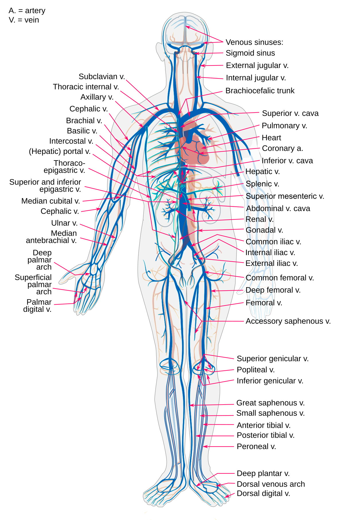
Provides blood to the Circumflex artery Runs to the posterior part of the heart, and it provides blood to the left atrium and left ventricle Left marginal artery Runs on the. Veins return blood to the heart from all the organs of the body The large veins parallel the large arteries and often share the same name, but the pathways of the venous system are more difficult to trace than those of the arteries Many unnamed small veins form irregular networks and connect with the large veins. The coronary sinus is a vein on the posterior side of the heart that returns deoxygenated blood from the myocardium to the vena cava Hepatic Portal Circulation The veins of the stomach and intestines perform a unique function instead of carrying blood directly back to the heart, they carry blood to the liver through the hepatic portal vein.
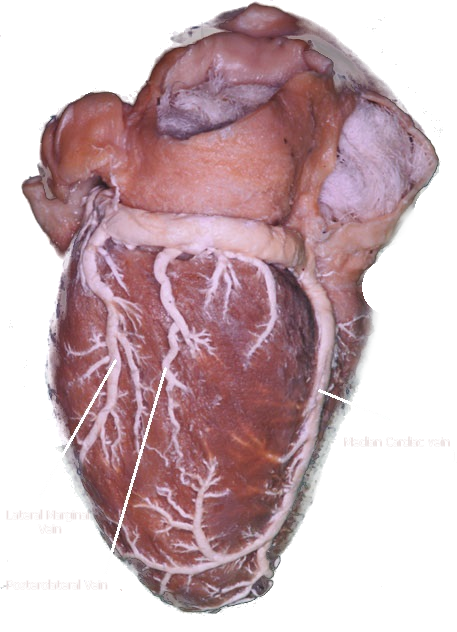
Start studying Major cardiac veins Learn vocabulary, terms, and more with flashcards, games, and other study tools. Robert H Anderson, in Paediatric Cardiology (Third Edition), 10 The Coronary Veins The venous return from the heart is, for the most part, collected by the major cardiac veins, which run alongside the coronary arteries in the interventricular and atrioventricular grooves. Introduction to the Coronary Veins After flowing through the myocardium, most (80%) of the oxygendepleted blood is returned to the right atrium by several prominent veins that run along the surface of the heart (= epicardial veins) Anterior Veins Draining blood from the anterior ventricles is the great cardiac vein.
Major veins A vein is defined as a vessel that conducts blood from the periphery to the heart All veins carry deoxygenated blood–except for the pulmonary vein The largest veins are the superior and inferior vena cava, and both drain directly into the right atrium of the heart All veins of the systemic circulation eventually drain back. Left coronary artery Originates from left posterior aortic sinus;. The heart also has its own blood supply, the cardiac arteries that provide tissue oxygenation to the heart as the blood within the heart is not used for oxygenation by the heart Cardiac Histology The heart is enclosed in a doublewalled protective membrane called the pericardium, which is a mesothelium tissue of the thoracic cavity.
It is presented below and is used consistently throughout 1 Vena cordis sinistra – the left cardiac vein (LCV) 2 Vena caudalis major – the major caudal vein (MCV) 3 Venae caudales minores – minor caudal veins (MiCV) 4 Vena cordis dextra – the right cardiac vein (RCV) 5 Venae cordis craniales –. This online quiz is called The major cardiac veins. The cardiac veins may be divided into three groups The largest system (Coronary Sinus and it tributaries), which collects a major amount of venous blood from the left ventricle and ends with the coronary sinus, opens into the right atrium;.
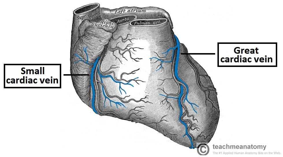
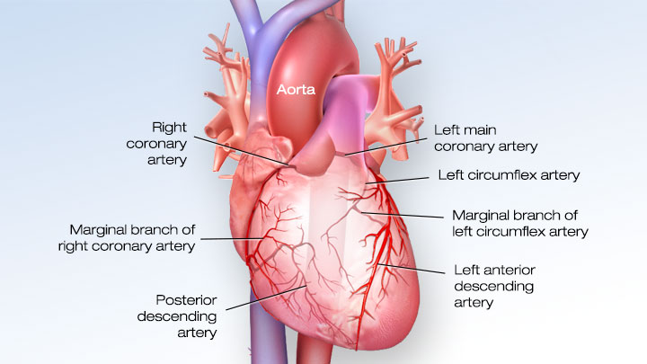
The second system (Anterior Right Ventricular veins) which gathers venous blood from the right twothirds of the right ventricle and ends in the right atrium. Two major coronary arteries branch off from the aorta near the point where the aorta and the left ventricle meet Right coronary artery supplies the right atrium and right ventricle with blood. All the veins of the heart (except for those that drain directly into the myocardium) drain into the coronary sinus There are three major veins The great cardiac vein drains the territory of the LAD and circumflex, the middle cardiac vein drains the region of the PDA and other posterior ventricular vessels, and the small cardiac vein drains the anterior wall of the right ventricle and right atrium.
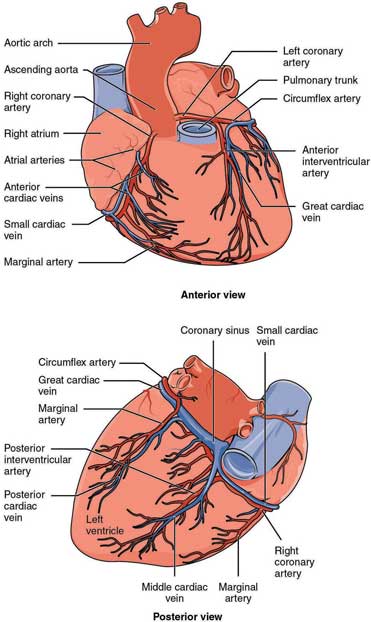
All 12 hearts with unidentifiable Thebesian veins had venous collaterals from the right ventricle (RV) to the major cardiac veins Epicardial veins extended to the proximal, middle, and distal thirds of the RV in 71%, 23%, and 6%, respectively CONCLUSION In ccTGA, the ventricular venous anatomy is abnormal and follows the morphologic RV. Most of the blood of the coronary veins returns through the coronary sinus The anatomy of the veins of the heart is very variable, but generally it is formed by the following veins heart veins that go into the coronary sinus the great cardiac vein, the middle cardiac vein, the small cardiac vein, the posterior vein of the left ventricle, and the vein of Marshall Heart veins that go directly to the right atrium the anterior cardiac veins, the smallest cardiac veins (Thebesian veins). The major cardiac veins in mice do not lie parallel to the branches of the coronary arteries, the latter lying intramurally The results presented above are markedly different not just from those of the human anatomy but also from well‐established descriptions of the heart venous system in larger animals,.
The middle cardiac veins and anterior interventricular veins were typically the longest (1297 and 1255 mm, respectively) and most tortuous veins (137 and 135) These 2 interventricular veins also had the most branches (68 and 99) and largest ostial long (57 and 54 mm) and short (41 and 45mm) diameters on average. We were talking about the heart again in this week's teaching, so here's an overview of the coronary arteries and cardiac veins. The coronary veins have a macroscopic disposition different from that of the coronary arteries and show many more variations Recent anatomic classification divides the cardiac veins into two main groups tributaries of the greater CVS and tributaries of the lesser CVS, which consists of the thebesian vessels (5–7).
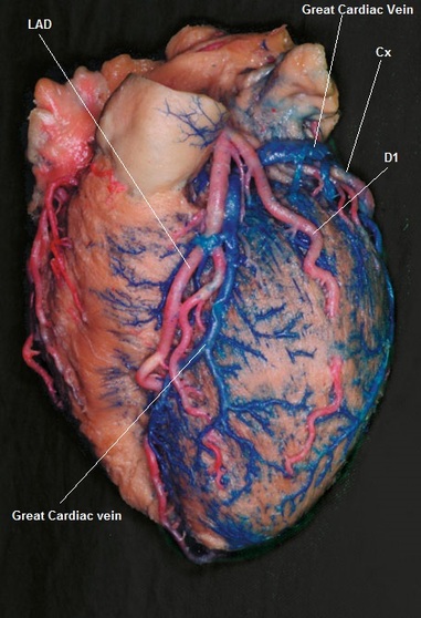
Murine cardiac veins consist of several principal branches (with large diameters), the distal parts of which are located in the subepicardium We have described the major branches of the left atrial veins, the vein of the left ventricle, the caudal veins, the vein of the right ventricle and the conal veins. As the great cardiac vein descends the atrioventricular groove, the great cardiac vein receives small tributaries (typically including a left marginal vein from the lateral wall or a left posterior vein draining the inferolateral wall) before joining the coronary sinus at the base of the heart (Fig 13). (2) the anterior cardiac veins, which primarily drain the anterior regions of the RV and the right cardiac border, ending principally in the RA;.
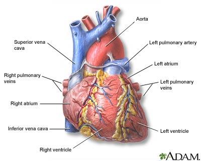
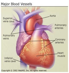
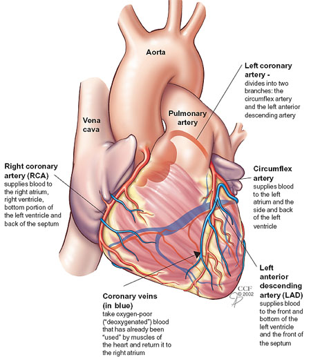
Pulmonary veins bring oxygenrich blood back to the heart from the lungs Right coronary artery (RCA) supplies blood to the right atrium, right ventricle, bottom portion of the left ventricle and back of the septum Inside the Heart The heart is a fourchambered, hollow organ. The coronary veins have a macroscopic disposition different from that of the coronary arteries and show many more variations Recent anatomic classification divides the cardiac veins into two main groups tributaries of the greater CVS and tributaries of the lesser CVS, which consists of the thebesian vessels (5–7). BCardiac veins follow same path as arteries include igreat cardiac vein runs with LAD iimiddle cardiac vein follows PDA iiismall cardiac vein runs with RCA ivoblique vein follows post part of LA 2anterior cardiac veins arise on ant surface of RV;.
And (3) the Thebesian venous network (venae cordis minimae), which is the smaller cardiac venous system and is composed of small venous branches made primarily of endothelial cells that are continuous with the lining. Major branches left anterior descending, left circumflex;. These are Brachiocephalic trunk Left common carotid artery Left subclavian artery.
Major branches left anterior descending, left circumflex;. This is a free printable worksheet in PDF format and holds a printable version of the quiz The major cardiac veins By printing out this quiz and taking it with pen and paper creates for a good variation to only playing it online. (Great cardiac vein labeled at center left) Pulmonary vessels, seen in a dorsal view of the heart and lungs The lungs have been pulled away from the median line, and a part of the right lung has been cut away to display the airducts and bloodvessels (great coronary vein labeled at center bottom).
Coronary veins Great, middle, small and oblique cardiac veins drain into the coronary sinus then into the right atrium. We were talking about the heart again in this week's teaching, so here's an overview of the coronary arteries and cardiac veins. Cardiac Veins Blood travels from the subendocardium into the thebesian veins, which are small tributaries running throughout the myocardiumThese in turn drain into larger veins that empty into the coronary sinus The coronary sinus is the main vein of the heart, located on the posterior surface in the coronary sulcus, which runs between the left atrium and left ventricle.
The structure and function of the heart, arteries, veins, and capillaries is vital for the circulatory system to work The overall function of the circulatory system is to transport blood and lymph around the body In doing so it delivers oxygen and nutrients to the body, removes waste products from the body, is involved in the. The great cardiac vein (left coronary vein) begins at the apex of the heart and ascends along the anterior longitudinal sulcus to the base of the ventricles It then curves around the left margin of the heart to reach the posterior surface. Major branches sinoatrial nodal, posterior descending, AV nodal, marginal;.
Drain directly into 4 chambers of the heart. Left coronary artery Originates from left posterior aortic sinus;. The structure and function of the heart, arteries, veins, and capillaries is vital for the circulatory system to work The overall function of the circulatory system is to transport blood and lymph around the body In doing so it delivers oxygen and nutrients to the body, removes waste products from the body, is involved in the.
Major branches sinoatrial nodal, posterior descending, AV nodal, marginal;. Overview Cardiac catheterization (kathuhturihZAYshun) is a procedure used to diagnose and treat certain cardiovascular conditions During cardiac catheterization, a long thin tube called a catheter is inserted in an artery or vein in your groin, neck or arm and threaded through your blood vessels to your heart. Drain into RA 3thebesian veins smallest;.
Representative Results Table 2presents the median anatomical parameters for the major cardiac veins for 42 human heart specimens All heart specimens contained one posterior interventricular vein (PIV) and anterior interventricular vein (AIV). Answer and Explanation The coronary arteries and cardiac veins are often found together in the grooves of the heart The left coronary artery splits into the left interventricular artery and the. There were 28 hearts with at least 1 Thebesian vein with an ostial opening >1 mm All 12 hearts with unidentifiable Thebesian veins had venous collaterals from the right ventricle (RV) to the major cardiac veins Epicardial veins extended to the proximal, middle, and distal thirds of the RV in 71%, 23%, and 6%, respectively.
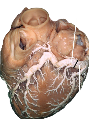
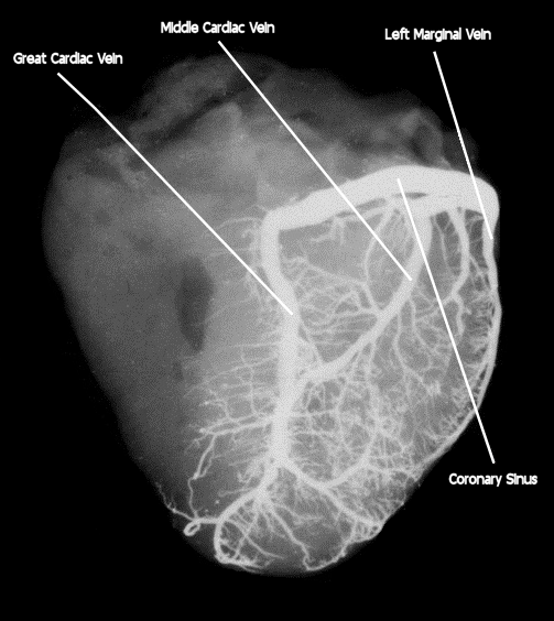
The CS originates at its ostium within the right atrium and extends distally to the valve of Vieussens, where it receives the great cardiac vein Other major tributaries include the left obtuse marginal vein, the posterior left ventricular vein (PCV), the middle cardiac vein (MCV), and the right coronary vein, also known as the small cardiac. The left and right coronary arteries branch off from the aorta and provide blood to the left and right sides of the heart The coronary sinus is a vein on the posterior side of the heart that returns deoxygenated blood from the myocardium to the vena cava Hepatic Portal Circulation. Your venous system is a network of veins that carry blood back to your heart from other organs We’ll explain the basic structure of a vein before diving into different types of veins and their.
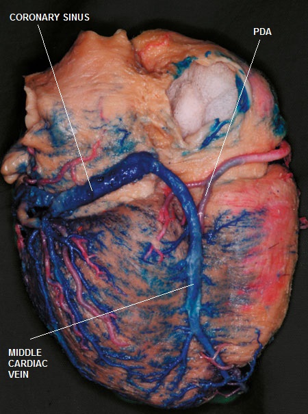
Middle Cardiac Vein The middle cardiac vein, also referred to as the posterior interventricular vein or more correctly, the inferior interventricular vein, is a major coronary vein that typically originates near the apex and usually ascends in or very near to the posterior interventricular sulcus 4,6,7. Share your videos with friends, family, and the world. Major veins A vein is defined as a vessel that conducts blood from the periphery to the heart All veins carry deoxygenated blood–except for the pulmonary vein The largest veins are the superior and inferior vena cava, and both drain directly into the right atrium of the heart All veins of the systemic circulation eventually drain back.
The coronary sinus is formed by three major veins of the heart, the great, middle and small cardiac veins It is approximately 25 cms in length and lies in the atrioventricular groove on the inferior wall of the heart, anterior to the atria, and posterior to the ventricles. Veins return blood to the heart from all the organs of the body The large veins parallel the large arteries and often share the same name, but the pathways of the venous system are more difficult to trace than those of the arteries Many unnamed small veins form irregular networks and connect with the large veins. Coronary arteries supply blood to the heart muscle Like all other tissues in the body, the heart muscle needs oxygenrich blood to function Also, oxygendepleted blood must be carried away The coronary arteries wrap around the outside of the heart Small branches dive into the heart muscle to.
Vascular disease is any abnormal condition of your blood vessels (arteries and veins) Learn more about the vascular disease types, causes, and treatment. Heart blockage is a term commonly used by patients referring to coronary artery disease, a buildup of plaque causing narrowing of the arteries that supply the heart muscle with blood This heart blockage, if severe enough, can prevent the muscle from getting the blood it needs to function, especially at times when more blood flow is required. Two major coronary arteries branch off from the aorta near the point where the aorta and the left ventricle meet Right coronary artery supplies the right atrium and right ventricle with blood.
Coronary veins Great, middle, small and oblique cardiac veins drain into the coronary sinus then into the right atrium. Share your videos with friends, family, and the world. Most cardiac veins collect and return blood to the right atrium through the coronary sinus;.
The myocardium drains mainly by three groups of veins (1) the CS and its tributaries, which return blood from almost the whole heart;. Anatomy of the major arteries and veins 3 Anatomy of the heart, the pericardium and valves – FROM CICM 3 Coronary artery anatomy 3 Anatomy of excitatory and conductive elements MAKEUP 4 Electrical properties of the heart 5 Ionic basis of automaticity the normal and abnormal processes of cardiac excitation 5 Pacemaker action potential 5. Start studying Major cardiac veins Learn vocabulary, terms, and more with flashcards, games, and other study tools.
Vascular disease is any abnormal condition of your blood vessels (arteries and veins) Learn more about the vascular disease types, causes, and treatment. Overview Cardiac catheterization (kathuhturihZAYshun) is a procedure used to diagnose and treat certain cardiovascular conditions During cardiac catheterization, a long thin tube called a catheter is inserted in an artery or vein in your groin, neck or arm and threaded through your blood vessels to your heart. Methods Pathologic cardiac specimens from patients with ccTGA were identified from the Mayo Clinic pathology database Coronary sinus (CS) anatomy and distances from the CS ostium to the major cardiac veins were evaluated Thebesian veins with ostial openings >1 mm, epicardial veins, and venous collaterals were also quantified.
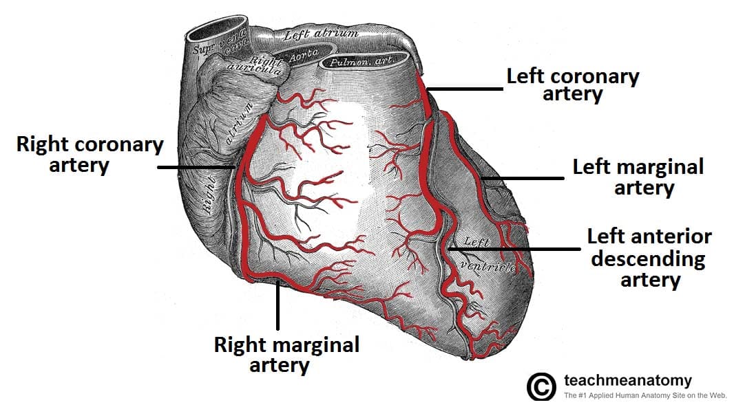
Vasculature Of The Heart Teachmeanatomy

Functional Anatomy Of The Cardiovascular System Clinical Gate

Carolyn S Jargon Free Patient Friendly Glossary Of Weird Cardiology Terms Heart Sisters
Major Cardiac Veins のギャラリー
Cardiac Veins Cthsurgery Com
Www Studocu Com En Au Document University Of Melbourne Human Structure And Function Lecture Notes Cvs2 Coronary Circulation And Great Vessels View

Coronary Sinus Dr S Venkatesan Md

Anatomy Of The Heart Coronary Circulation Medical Anatomy Medical Textbooks
Cardiac Veins Cthsurgery Com

Overview Of The Heart Anatomy A Illustration Of The External Anatomy Download Scientific Diagram
Lecture 4 Heart Anatomy
Anatomy Thorax Coronary Sinus Article
Cardiac Veins Cthsurgery Com
Coronary Veins Cardiac Veins

Lab 1 Major Cardiac Veins Diagram Quizlet
Q Tbn And9gcs4vuhnoufbevgxb6zoslqk V2sb8kvoeexlpglufaocnbfiuzs Usqp Cau
Coronary Artery Disease Causes Symptoms Diagnosis Treatments

Anatomy Of The Cardiovascular System 3
Blood Flow Of The Heart
Q Tbn And9gcqjawasd7zbmwjoiajkffajronk0zu3kcromxby3hfye2upfysh Usqp Cau
Heart Front View Medlineplus Medical Encyclopedia Image

Coronary Veins Cardiac Veins
Lab Guide Sites Uci

Heart Anatomy Anatomy And Physiology

Cardiac Veins An Anatomical Review Sciencedirect

Major Blood Vessels Leading To The Heart Superior Vena Cava Inferior Vena Cava Coronary Sinus Video Lesson Transcript Study Com
Great Cardiac Vein Wikipedia
Anatomy And Circulation Of The Heart

Anatomy Of The Cardiovascular System 3
Coronary Arteries How It Works Images
Illustrations Of The Blood Vessels

Quotes About Blood And Veins Quotes

Blood Vessels Biology For Majors Ii
Your Heart Blood Vessels

Coronary System Tutorial What Is The Coronary System

Cardiovascular System Physiopedia

Coronary Veins Cardiac Veins

Major Cardiac Veins Part 1 Diagram Quizlet

Basic Anatomy Of The Human Heart The Cardio Research Web Project
:background_color(FFFFFF):format(jpeg)/images/library/13977/Coronary_vessels_cardiac_veins.png)
Coronary Arteries And Cardiac Veins Anatomy And Branches Kenhub

Solved 2 Label The Major Arteries And Veins On The Poste Chegg Com

Mammalian Heart And Blood Vessels Boundless Biology
Cardiac Surgery Basicmedical Key

Coronary Circulation Of The Heart Bioscience Notes
Coronary Circulation Wikipedia
Vasculature Of The Heart Teachmeanatomy

The Heart And Arteries Veterian Key
Anatomy And Physiology Of The Cardiovascular System Thoracic Key
Cardiac Veins Cthsurgery Com
Illustrations Of The Heart Showing The Coronary Venous System And The Download Scientific Diagram

Overview Of The Heart Anatomy A Illustration Of The External Anatomy Download Scientific Diagram

Anatomy 2 Study Guide 2 Biol8 Studocu

Major Blood Vessels Of The Heart
Chapter 18 The Cardiovascular System The Heart Part B Ppt Download

Heart Major Coronary Arteries Cardiac Veins Flashcards Quizlet

The Coronary Arteries And Major Veins Of The Heart Anterior And Inferior Views Biology Forums Gallery
Coronary Circulation Course Hero

Heart Anatomy Anatomy And Physiology

Coronary Veins Cardiac Veins
Cardiac Veins Cthsurgery Com
Vein Wikipedia
Circulatory System

The Heart Part 1 Slides By Vince Austin And W Rose Ppt Download

Seer Training Structure Of The Heart

Major Differences Difference Between Artery And Vein
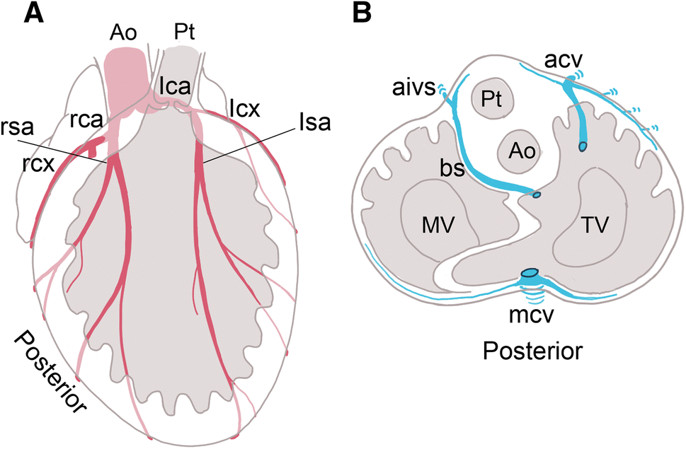
Figure 3 Anatomy Of The Coronary Artery And Cardiac Vein In The Quail Ventricle Patterns Are Distinct From Those In Mouse And Human Hearts Springerlink
Cardiac Veins Cthsurgery Com

Anatomy And Cell Biology 3319 Lecture Notes Fall 17 Lecture 34 Small Cardiac Vein Femoral Triangle Left Coronary Artery

Heart Anatomy Anatomy And Physiology
Cardiovascular Media Library Watch Learn Live

Great Cardiac Vein An Overview Sciencedirect Topics

12 December 08 Dote Anatomy Topics

Venous Drainage Of The Heart Skudra Net Human Heart Anatomy Drainage Heart Anatomy

Pedi Cardiology Anatomy Coronary Veins Coronary Arteries Coronary Arteries Arteries Anatomy Arteries And Veins
Body Anatomy Upper Extremity Vessels The Hand Society
Http Www Gmch Gov In Sites Default Files Documents Thorax Heart Blood Supply Innervation Pdf

X Anlwjmsfut M
Cardiac Veins Cthsurgery Com

Cardiovascular System Human Veins Arteries Heart

Overview Of Coronary Artery Disease Cad Heart And Blood Vessel Disorders Msd Manual Consumer Version
Link Springer Com Content Pdf 10 1007 2f978 3 642 4 6 Pdf

Cardiac Veins An Anatomical Review Sciencedirect
3
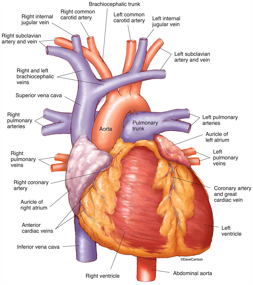
Cardiovascular System Carlson Stock Art
/vascular-system-veins-56c87fa03df78cfb378b3e7c.jpg)
What Is A Vein Definition Types And Illustration
Q Tbn And9gcs4vuhnoufbevgxb6zoslqk V2sb8kvoeexlpglufaocnbfiuzs Usqp Cau

Small Cardiac Vein Wikipedia
Http Ksumsc Com Download Center Archive 1st 437 4 Cardiovascular block Teamwork Anatomy L4 anatomy of the arterial supply and venous drainage of the heart Pdf

How The Heart Works Nhlbi Nih
:watermark(/images/watermark_5000_10percent.png,0,0,0):watermark(/images/logo_url.png,-10,-10,0):format(jpeg)/images/atlas_overview_image/265/I5tZeGiyLifEWKeirVrJ0Q_coronary-arteries-and-cardiac-veins_english.jpg)
Coronary Arteries And Cardiac Veins Anatomy And Branches Kenhub
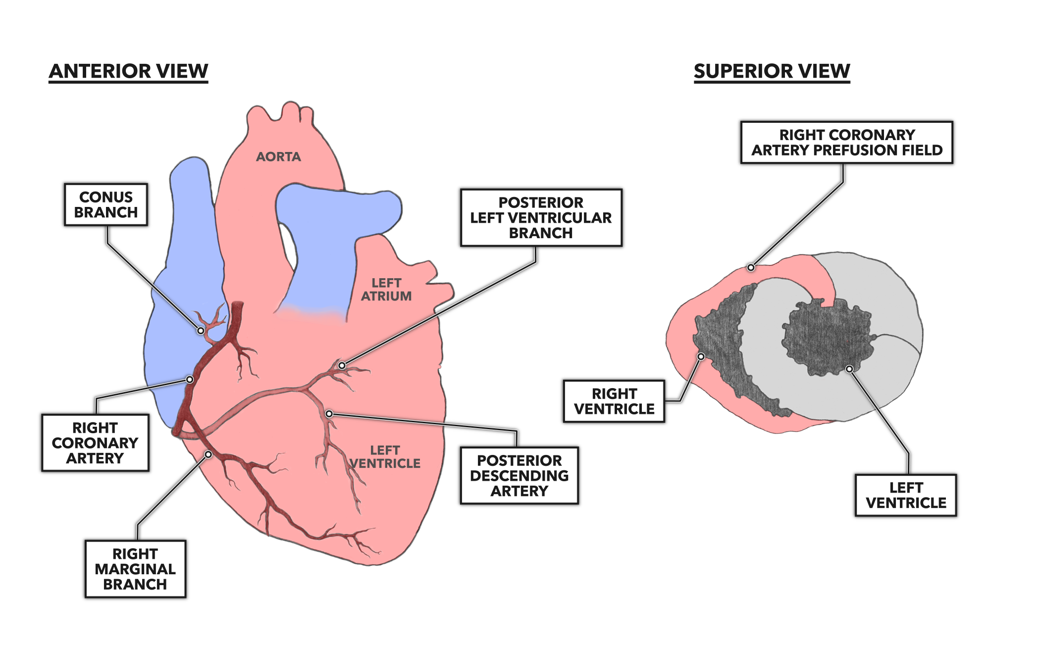
Crossfit The Heart Part 7 Coronary Circulation
:background_color(FFFFFF):format(jpeg)/images/library/12591/Heart.png)
Coronary Arteries And Cardiac Veins Anatomy And Branches Kenhub

The Heart
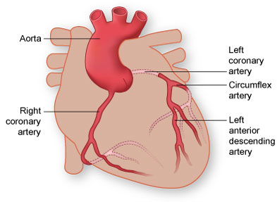
Coronary Arteries Texas Heart Institute

Coronary Veins Cardiac Veins

109 Single Coronary Type R2a Dr Buchanan S Cardiology Library Vin
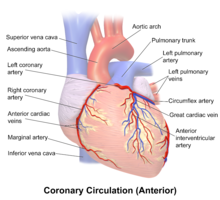
Coronary Arteries Wikipedia

Coronary Circulation Anatomy Demonstration Video Medchrometube

Know Ur Heart The Coronary Circulation Coronary Circulation Arteries And Veins Coronary Arteries
:background_color(FFFFFF):format(jpeg)/images/article/en/blood-supply-of-the-heart/MJoHBHSCRsCRGLekSkbMdA_Left_coronary_artery.png)
Coronary Arteries And Cardiac Veins Anatomy And Branches Kenhub
The Anatomy Of The Human Heart Dummies

Great Cardiac Vein An Overview Sciencedirect Topics

Anatomy Of The Heart And Major Coronary Vessels In Anterior Left And Download Scientific Diagram
Anatomy And Function Of The Coronary Arteries Johns Hopkins Medicine
Blood Supply To The Heart Thoracic Key


